Device Closure Procedure | ASD | VSD | PDA
Saraswati Dayal’s Motiramani Heart Clinic is one of the most trusted Device Closure Procedure ASD, VSD, And PDA Clinics in Shankar Nagar, Raipur Chhattisgarh, India.
About Device Closure Procedure ASD, VSD, PDA
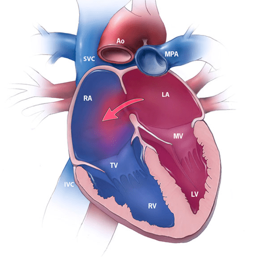
Device Closure is used to close a defect or an opening between the right and left sides of the heart. A heart has four chambers. The upper two chambers are called at Atria. Atria are separated by a wall which is called Atrial Septum. The lower chambers are known as Ventricles- Right and left. Pulmonary Veins supply Oxygenated blood to the Left Atrium. This left atrium, in turn, sends this oxygenated blood to the Left Ventricle through Mitral Valve, for circulation of oxygenated blood in the body.
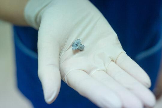
The walls of the heart are well separated maintaining a good circulation system. But in some congenital heart defect situations, if there are any holes on the heart, either in the atrial wall or the ventricle wall, this can result in disturbed blood circulation resulting in performance failure of the heart. If the holes are particularly small, they can be filled up surgically, with some metal parts. This concept is known as DEVICE CLOSURE. So, in short, in Device Closure, there is a metal device that is surgically placed in the place where there is a whole in a heart, in order to maintain the heart’s performance.
What is Device Closure Procedure?
The device closure procedure used as a reparative surgery is known as transcatheter closure, although it is done for a reparable hole and this is not the only treatment for hole closures. When compared to other surgeries, it is a less invasive surgery.
DURING THE SURGERY:
Before the surgery starts, a patient is administered with IV and intravenous medicines are started. The person has leads attached to his chest for ECG. The person’s groin/arm area is cleaned and a plastic thin tube called a catheter will be inserted through that area. Meanwhile, the patient is given sedatives for relaxation purposes and local anesthesia, but they will be awake and conscious throughout the procedure.
A contrast dye would be injected in the arteries so as to get an idea of the heart chambers and the size of the hole. If required, an echocardiogram or transoesophageal echocardiogram is also taken.
After the size of the hole is determined, a special catheter is administered in the arteries which carry a small metallic mesh-like closing device. This device is then fitted in the hole and then released by the catheter. This catheter is then taken out, eventually, heart tissue develops on the closure device, thus making it a part of the heart itself.
POST SURGERY:
When the catheter is taken out, suturing of the area is done or heavy bandaging, as required. The patient is kept in the hospital for a day or two. The patient can resume their routine a week after the doctor’s advice. They are advised to maintain a healthy lifestyle which includes exercising, maintaining a good diet, and regular cardiac checkups.
ASD Device Closure Procedures
An ASD closure device (atrial septal defect) is an opening in the membrane between two chambers of the heart. Right Atrium consists of Deoxygenated blood and through that, it is sent to the lungs for purification after passing through the Right Ventricle. In Atrial Septal Disorder, there is exists a small hole in the wall separating the atria, called the atrial septum. Due to this, Oxygen-rich blood leaks from the left atrium to the right side resulting in the reverse flow of blood. As a result of this, right atria gets loaded with extra work as the oxygenated and deoxygenated blood gets mixed up, resulting in more pressure on the right atrium to send more blood to the lungs.
The typical symptoms include Heart murmurs as there is more blood flowing through the pulmonary vein shortness of breath, easy fatigue after exercise, heart palpitations, etc. These symptoms can arise early in childhood & they may also reflect in adulthood.
Patients with ASD are always suggested for a device closure surgery before symptoms get worsened & irreversible changes happen in the heart.
Right Atrium consists of Deoxygenated blood and through that, it is sent to the lungs for purification after passing through the Right Ventricle. In Atrial Septal Disorder, there is exists a small hole in the wall separating the atria, called as the atrial septum. Due to this, Oxygen rich blood leaks from the left atrium to the right side resulting in reverse flow of blood. As a result of this, right atria gets loaded with extra work as oxygenated and deoxygenated blood gets mixed up, resulting in more pressure on the right atrium to send more blood to the lungs.
There are four types of ASD which are as follows:
- Secundum ASD: It’s a condition where a hole occurs in the middle part of the wall between the atria (atrial septum). This is the most common type of ASD.
- Primum ASD: This defect arises in the lower part of the atrial septum and might be coupled with other congenital heart issues.
- Sinus venosus ASD: This is a rare defect. It usually occurs in the upper part of the atrial septum and this also is often coupled with other congenital heart issues.
- Coronary sinus ASD: In this rare defect, part of the wall between the coronary sinus — which is part of the vein system of the heart — and the left atrium is absent.
The typical symptoms include, Heart murmurs as there is more blood flowing through the pulmonary vein; shortness of breath, easy fatigue after exercise, heart palpitations, etc. These symptoms can arise early in childhood & they may also reflect in adulthood.
Small defects may never cause a serious issue, although larger defects may create some complications such as Right side heart failure, an increase in the risk of a stroke, shortened life span, pulmonary hypertension, Eisenmenger Syndrome etc.
Patients with ASD are always suggested for a device closure surgery, before symptoms get worsened & irreversible changes happen in the heart.
VSD Device Closure Procedure
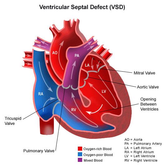
VSD device closure is an opening in the wall (septum) dividing the two lower chambers of the heart (ventricles). Normally, this wall closes before birth. The lower two chambers of the heart are called Ventricles. These are separated by a membrane, called the Ventricular Septum, which prevents the leakage of the oxygenated blood from the left ventricle to the right ventricle containing deoxygenated blood. This deoxygenated blood from the right ventricle is then sent to the lungs for purification.
In VSD device closure, there exists a hole in the ventricular septum. Because of this, the oxygenated blood gets mixed up with the deoxygenated blood as the reversal of the blood flow takes place from left to right. As a result, there is excessive blood pumping to the lungs & the heart gets an overload to supply oxygenated blood to the body.
This defective hole varies in size. It can be as small as a pinpoint and in extreme cases, there can be no ventricular septum, meaning a single large ventricle with mixed up oxygenated and deoxygenated blood.
Typical symptoms can manifest early in childhood or won’t show up until adulthood. The most common symptoms are Shortness of breath, poor eating leading to not gaining weight, easy tiring, heart murmurs and many others.
If the hole is small, it can be fixed with a device closure which involves heart catheterization. It involves little risk or no risk at all depending upon the complexity.
The operation has been widely used & successfully majority of the times, about 6 percent of patients have required a re-operation to close small leaks that developed around the patch. It has been under observation that adults with closed VSDs and without other heart or lung issues can expect to live a normal life.
Within the first six months after device closure repair of a VSD, the person still has a little risk of endocarditis while the heart heals. Your cardiologist will advise you on how to protect yourself from this life-threatening condition.
PDA Device Closure Procedure
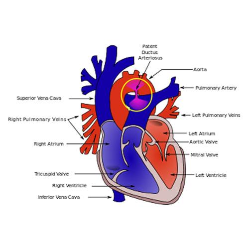
Patent means ‘open’. And ductus arteriosus is a normal blood vessel. So, in short, there exists an abnormal opening in a normal blood vessel. In a normal human heart, Aorta supplies oxygenated blood to the whole body & Pulmonary artery carries deoxygenated blood to the lungs. Both blood vessels are separated from each other. When a baby is in the womb, there’s a small duct that connects the aorta and the pulmonary artery, which is normal. This small duct is called as Ductus Arteriosus. It is because; lungs of a baby start working only when it is born. It keeps getting pure blood supply from the placental connection with its mother. When the baby gets birth, this ductus arteriosus gets closed within one-two days and in some conditions, up to one year. A defect gets detected when the opening doesn’t close and Aorta & pulmonary artery still has an open connection between them. Because of this, the oxygen-rich blood from aorta gets leaked into the pulmonary artery resulting in an overpressure to the lungs. The long-term prognosis for a patient undergoing PDA device closure either by surgery or device closure is outstanding.
The exact cause of PDA is unknown, yet genetics is responsible. PDA is more common in premature babies & often accompanied by Down’s syndrome and some genetic abnormalities. Usually this is a fairly straightforward surgery.
General signs & symptoms of PDA
Babies with a large PDA might have symptoms such as:
- A bounding pulse
- Fast breathing
- Not feeding well
- Shortness of breath
- Sweating while feeding
The patent ductus arteriosus is diagnosed with chest X-ray & EKG. If required, a contrast dye is also administered in a catheterization. This ductus arteriosus is either sewed or closed by a PDA Device Closure. In premature and small babies typically surgery is used to close a PDA.